


Knee Replacement Pictures: What Your Post-Op Images Show
Most people who search “knee replacement pictures” aren’t looking for surgical photography — they want to understand the X-rays and scans their own surgical team will use to check their new joint. Those clinical images are what actually matter for your recovery, and they follow a predictable schedule that most patients are never walked through in advance. Expect between three and four rounds of imaging in your first year: immediately after surgery to confirm placement, around six weeks to check early healing, and again at three to six months to verify the implant has bonded properly with your bone.
What Do Knee Replacement Pictures Show After Surgery?
Knee replacement pictures taken after surgery are almost always X-rays, and occasionally CT scans, that show the position of your new implant against your femur and tibia. They confirm three things: whether the components are aligned correctly, whether the bone is bonding to the implant surface as expected, and whether there are any early warning signs a surgeon would want to watch. None of this is about how the surgery looked from the outside — it’s diagnostic, not descriptive.
A standard post-op X-ray is taken from at least two angles, front-on and side-on, so the surgical team can judge alignment in three dimensions rather than guessing from a flat image. Radiologists and orthopaedic surgeons read these differently to how a patient would: a shadow or line that looks alarming to an untrained eye is often exactly what a correctly seated implant is supposed to look like. If you’d rather see the surgical side of the process — the incision, the implant components themselves, the operating theatre view — our detailed breakdown of what knee replacement photos actually show covers that separately.
- Anteroposterior (front-facing) X-ray — checks left-right alignment of the components
- Lateral (side-on) X-ray — checks the implant’s tilt and depth
- Occasional CT scan — used only when a standard X-ray raises a question that needs 3D detail to resolve
Key Takeaways
- Knee replacement pictures that matter clinically are X-rays and occasional CT scans, not surgical photography — they check implant alignment, bone bonding, and early complications.
- Post-op imaging follows a predictable schedule: immediately after surgery, around six weeks, and again at three to six months.
- Findings like small bone-implant gaps or metal artefacts on an X-ray are usually normal and expected, not a sign that anything has gone wrong.
- 3D laparoscopic imaging during surgery gives the surgical team real-time visualisation of the joint that standard 2D imaging can’t match, improving how precisely the implant is positioned in the first place.
- Patients who understand what their own images show tend to worry less and stick more closely to their physiotherapy plan.
When Are Knee Replacement Images Taken After Surgery?
Post-op imaging for a total or partial knee replacement generally happens at three points: within 24 hours of surgery, around the six-week mark, and again between three and six months out. Each stage is checking for something different, so skipping the sequence isn’t an option even if you feel fine.
The first X-ray, taken before you leave the hospital, is purely a placement check — it confirms the implant sits where the surgeon intended before you start putting weight through it. The six-week image looks for early bone response around the implant, which matters most if you’re recovering from a full total knee replacement rather than a smaller partial procedure. By three to six months, the goal shifts to confirming stable integration, the point at which most surgeons clear patients for higher-impact activity.
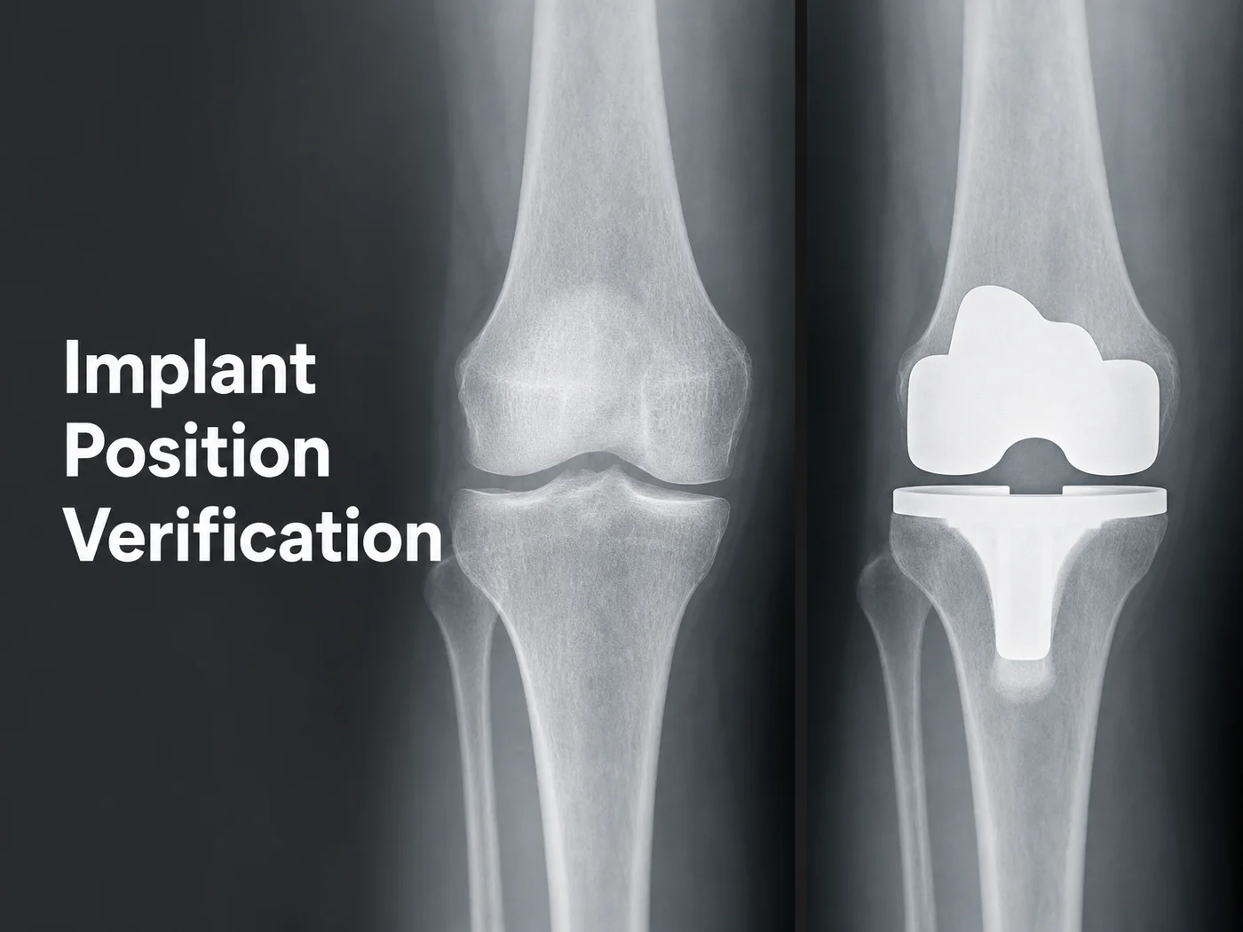
A simplified view of what an X-ray checks at each stage — alignment first, then bone integration over time.
Some patients need an unscheduled image sooner than the standard timeline, usually because of swelling that doesn’t settle, a specific pain pattern, or a fall during early recovery. That’s not a sign the surgery went wrong; it’s the same caution any orthopaedic team applies when something falls outside the expected pattern.
Normal Findings on Post-Op Images Are More Common Than You Think
Patients who look up their own scans online often panic at things that are routine. A thin dark line at the edge of the implant, sometimes called a lucent line, can simply reflect how bone remodels around metal over the first few months rather than a loosening implant. Metal artefacts — the starburst-like distortion around the implant on an X-ray or CT — are a property of imaging metal, not a defect in the surgery itself.
Worth knowing: Radiologists expect some visual “noise” around a metal implant on every scan. It’s the pattern of change over multiple images, not any single picture, that tells a surgeon whether healing is on track.
Your surgical team reads these images alongside your symptoms, your walking pattern, and your physiotherapy progress — never the image in isolation. That combined view is exactly why a picture that looks concerning to you can be entirely unremarkable to the person trained to read it.
Why Advanced Imaging Matters for Your Knee Replacement
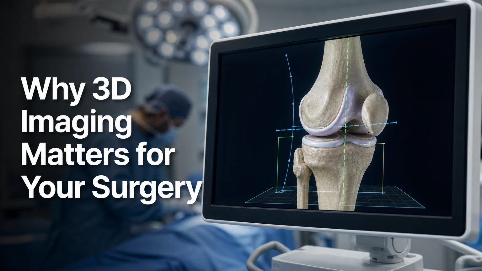
Real-time 3D visualisation during surgery, rather than a flat 2D view, is what shapes how accurately the implant is placed in the first place.
The imaging that matters most for a good long-term result actually happens during surgery, not after it — and this is where the technology used makes a measurable difference. At KCM Clinic, total and partial knee replacement is performed using 3D laparoscopic-assisted imaging, which gives the surgical team continuous depth perception of the joint as they work, rather than the flat, two-dimensional view that standard surgical imaging provides.
Standard 2D visualisation asks a surgeon to judge depth and alignment largely from experience and repeated 2D reference points. 3D laparoscopic imaging removes much of that guesswork by rendering the joint’s true spatial geometry in real time, which is particularly valuable during bone resection and trial-implant fitting — the two steps where a small misjudgement has the biggest effect on how the post-op X-rays will look weeks later. Better intraoperative visualisation is one of the clearest ways a surgical team can reduce the risk of implant misalignment before it ever shows up on a follow-up scan.
This is also where post-op imaging and surgical technique connect directly: a well-positioned implant, confirmed by good 3D visualisation during the operation, is far more likely to produce unremarkable, reassuring images at your six-week and six-month checks.
Are Knee Replacement Pictures and X-Rays the Same Thing?
Not quite — “knee replacement pictures” is a broad search term covering everything from surgical photography to X-rays and 3D scans, while an X-ray is one specific diagnostic image type used to check implant position. Understanding the difference matters because each format answers a different question.
| Image type | What it shows | When it’s used |
|---|---|---|
| X-ray | Implant alignment, bone density, spacing | Immediate post-op, 6-week, 3–6 month checks |
| CT scan | 3D bone and implant detail | Only when an X-ray raises a question needing more depth |
| 3D intraoperative imaging | Real-time joint geometry during surgery | Throughout the procedure itself, not after |
| Surgical photography | The physical steps of the operation | Rarely used clinically; mostly educational |
If a search result shows you a graphic surgical photo, it’s almost certainly educational content, not something your own surgical team will generate or rely on. Your actual post-op images will be X-rays, and occasionally a CT scan if something needs a closer look.
Get a Clearer Picture With KCM Clinic
Understanding what your post-op images are checking for tends to settle far more nerves than the images themselves. KCM Clinic performs total and partial knee replacement using 3D laparoscopic-assisted imaging and named implant systems, with every procedure built into a single all-inclusive package for patients travelling from the UK and Canada.
Book a Free Consultation with KCM Clinic
Frequently Asked Questions
Are knee replacement pictures the same as X-rays?
Not exactly. “Knee replacement pictures” is a general search term that can mean surgical photography, X-rays, or scans, while an X-ray is a specific diagnostic image your surgical team uses to check implant alignment and bone healing after your operation.
How long after knee replacement will I get my first images?
Your first X-ray is usually taken within 24 hours of surgery, before you’re discharged, purely to confirm the implant is positioned correctly. Further images typically follow around six weeks and again at three to six months.
What if my knee replacement X-ray shows a gap next to the implant?
Small gaps or thin lucent lines next to an implant are common on early post-op X-rays and often reflect normal bone remodelling rather than a problem. Your surgical team compares this against your symptoms and prior images before deciding whether any follow-up is needed.
Does KCM Clinic use 3D imaging for every knee replacement?
Yes. KCM Clinic performs total and partial knee replacement with 3D laparoscopic-assisted imaging as standard, giving the surgical team real-time depth perception during the procedure rather than relying on flat 2D visualisation alone.