
Chondrocalcinosis Knee: What It Is and How It’s Treated
Physician-reviewed patient education from KCM Clinic — medically reviewed by Dr. hab. n. med. Stanisław Kwiek, Specialist Neurosurgeon, KCM Clinic.
Chondrocalcinosis knee findings are rarely the reason someone books an X-ray in the first place. Far more often, it’s the incidental finding — a faint line of calcium sitting inside the cartilage that a radiologist notices while looking for something else, and which then becomes the diagnosis nobody was expecting.
That’s not a failure of the healthcare system. It’s simply how this condition tends to surface, and it shapes almost everything about how it should be managed from that point on.
What Is Chondrocalcinosis Knee Damage and Why Does It Develop?
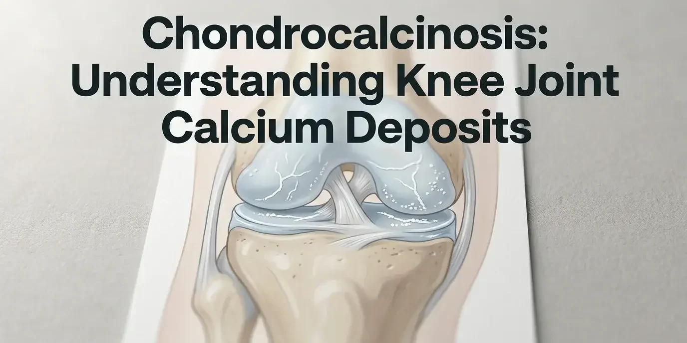
Chondrocalcinosis is the deposit of calcium pyrophosphate crystals within joint cartilage, most commonly seen on knee X-rays as a thin calcified line running through the meniscus or articular surface. It’s also known as calcium pyrophosphate deposition disease (CPPD), and when it flares acutely with pain and swelling, clinicians call it pseudogout — a name that reflects how closely the attack can mimic true gout.
The knee is the joint most frequently affected, followed by the wrist and shoulder. Age is the strongest risk factor: chondrocalcinosis is uncommon before 60 and becomes progressively more prevalent after that, though it can appear earlier in people with certain metabolic conditions such as haemochromatosis or hyperparathyroidism.
Not everyone with calcium deposits on an X-ray has symptoms. Many patients carry chondrocalcinosis for years without pain, discovered only when imaging is done for an unrelated reason.
The Crystals Behind the Condition
Calcium pyrophosphate crystals form when the body’s normal process for clearing pyrophosphate — a byproduct of cellular metabolism — breaks down within the cartilage matrix. Instead of being recycled, the compound binds with calcium and settles into microscopic deposits.

Calcium pyrophosphate crystals accumulate gradually within the cartilage matrix, often over years before any symptom appears.
These crystals are chemically distinct from the urate crystals that cause true gout, which is why pseudogout requires a different long-term management approach even though the acute attack looks nearly identical to a patient. Joint fluid analysis under polarised light microscopy is the definitive way to tell the two apart when an attack is severe enough to warrant it.
Chondrocalcinosis and Osteoarthritis Share a Complicated Relationship
The two conditions overlap enough that patients often ask whether they’re the same thing. They aren’t, but the relationship runs deeper than coincidence.
- Chondrocalcinosis is frequently found alongside existing osteoarthritis in the same knee
- Crystal deposits can accelerate cartilage breakdown once osteoarthritis is already present
- Some research suggests chondrocalcinosis may itself act as a risk factor for developing knee osteoarthritis over time
- The two conditions are graded and monitored separately, even when they coexist on the same scan
Worth knowing: A knee can have chondrocalcinosis without any osteoarthritis, and vice versa. When both appear together, treatment usually targets whichever is driving the current symptoms — not both indiscriminately.
This is one reason imaging alone rarely settles a treatment plan. A calcified meniscus on an X-ray tells a radiologist what’s there; it doesn’t tell a surgeon how much it’s contributing to the pain a patient is actually feeling.
Diagnosis Usually Starts with an X-Ray, Not a Symptom Pattern
Standard weight-bearing X-rays are typically the first step, since the calcified cartilage is often visible without any further imaging. Ultrasound can pick up crystal deposits that X-ray misses, particularly in early or mild cases, and is increasingly used alongside plain film.
MRI becomes useful when a clinician needs to assess soft tissue damage or rule out a meniscal tear happening independently of the calcification. Blood tests are sometimes ordered too, mainly to check for the metabolic conditions — thyroid disorders, haemochromatosis, elevated calcium — that can predispose someone to CPPD at a younger age than typical.
Medical advice is worth seeking when knee pain is accompanied by sudden swelling, warmth, or stiffness that limits movement, particularly if it recurs in flares rather than building gradually. A GP or rheumatologist can distinguish a pseudogout flare from other causes and start the right first-line treatment quickly.
Conservative Treatment Comes First — and Often Works for Years
For most patients, chondrocalcinosis is managed without surgery for a long time, sometimes indefinitely. NSAIDs remain the standard response to an acute flare, reducing both pain and the inflammation driving it. Rest and joint protection during a flare-up matter just as much as medication.
Between flares, a physiotherapist-guided strengthening programme helps stabilise the joint and can reduce how often attacks occur, though the specific exercises should be set by a clinician who’s examined the knee rather than followed generically. Intra-articular corticosteroid injections are often used for more frequent or stubborn flares, and low-dose colchicine is sometimes prescribed for patients with recurring episodes.
None of these treatments reverse the calcium deposits already present. What they do is manage symptoms and slow the cartilage damage that repeated inflammation can cause — which, for many patients, is enough to avoid surgery for years.
When Is Surgery Necessary for Chondrocalcinosis Knee Damage?
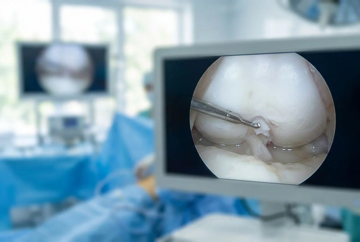
Arthroscopy accesses the joint through small ports guided by a camera, rather than the open incision traditional surgery requires.
Surgery for chondrocalcinosis becomes a serious consideration when conservative treatment stops controlling pain, or when crystal-driven cartilage damage has advanced enough to cause a mechanical block — locking, catching, or a persistent loss of range of motion. At that stage, the choice of surgical approach matters as much as the decision to operate at all.
At KCM Clinic, early-stage cartilage loss linked to chondrocalcinosis is addressed arthroscopically wherever the joint anatomy allows it, rather than defaulting to open surgery. Arthroscopic protocols access the joint through small ports rather than a large incision, and where cartilage or meniscal tissue needs support or repair, the surgical team uses joint-preservation implant systems from Lima and Zimmer Biomet — devices designed to stabilise or restore the affected structure without sacrificing healthy surrounding cartilage.
Arthroscopy vs Traditional Open Surgery vs Total Knee Replacement
Traditional open surgery still has a role, particularly where damage is extensive or affects multiple compartments of the knee. But for the earlier-stage presentations that chondrocalcinosis most often produces, the arthroscopic route is generally the less invasive first option.
| Approach | Access | Typical Hospital Stay | Best Suited For |
|---|---|---|---|
| Arthroscopic joint preservation | Small camera-guided ports | Often same day to one night | Early-to-moderate cartilage or meniscal damage, single compartment |
| Traditional open surgery | Full surgical incision | Several days | Extensive or multi-compartment joint damage |
| Total knee replacement | Full surgical incision, implant across the joint | Several days | End-stage joint destruction where preservation is no longer viable |
Patients whose cartilage loss has progressed past what preservation surgery can address may eventually need a total knee replacement, and those with damage confined to one compartment are sometimes better candidates for a partial knee replacement than a full one. Which pathway applies depends on imaging and, usually, a diagnostic arthroscopy first.
Recovery and Long-Term Outlook After Arthroscopic Treatment
Recovery from arthroscopic joint-preservation surgery is generally measured in weeks rather than months. Most patients bear weight within days, guided by a physiotherapy programme that typically starts before discharge and continues for several weeks afterward.
That timeline sits in contrast to recovery from a partial or total knee replacement, which involves a longer initial rehabilitation phase even though long-term outcomes for replacement can be excellent when it’s genuinely needed. Patients researching partial knee replacement recovery timelines should note that arthroscopic joint preservation, when it’s a viable option, is a meaningfully shorter path back to normal activity.
Long-term outcomes depend heavily on how much cartilage damage existed before treatment. Caught early, arthroscopic intervention can delay or avoid the need for replacement surgery for years. Left untreated through repeated flares, the joint damage that chondrocalcinosis contributes to can accelerate the timeline toward total knee replacement regardless of which country a patient is treated in.
What This Means If You’re Weighing Treatment from the UK or Canada
NHS knee replacement pathways for advanced arthritis typically involve a lengthy referral and waiting-list process, and elective surgery for a condition like chondrocalcinosis-related cartilage damage often sits well down that queue. That timeline is a major reason UK and Canadian patients start researching private options once conservative treatment stops holding.
Private knee replacement cost in the UK varies significantly by hospital, surgeon, and implant, and headline prices don’t always include the full pre- and post-operative care a joint procedure needs. KCM Clinic’s packages bundle the consultation, procedure, hospital stay, and physiotherapy into one all-inclusive arrangement, treated within the same EU Medical Devices Regulation framework that governs Germany and France. Before any joint procedure, patients also go through the kind of broader pre-operative health screening used across KCM’s surgical programmes, which can flag related concerns — including bone-density issues relevant to osteoporosis treatment — before they complicate recovery.
Quick recap
- Chondrocalcinosis (CPPD or pseudogout) is calcium crystal buildup in the cartilage, most common in the knee and typically diagnosed by X-ray, ultrasound, or MRI
- It’s distinct from osteoarthritis but frequently coexists with it, and each is managed on its own terms
- NSAIDs, rest, physiotherapy, and joint injections control most cases for years without surgery
- When surgery is needed, arthroscopic joint-preservation techniques using implants such as Lima and Zimmer Biomet systems offer a less invasive route than traditional open surgery for early-to-moderate cartilage damage
- UK and Canadian patients facing long NHS waits can access EU-standard arthroscopic care at KCM Clinic with bundled aftercare
Get Chondrocalcinosis Treatment Options at KCM Clinic
If conservative treatment for chondrocalcinosis is no longer holding, or you want to understand your surgical options before joint damage advances further, KCM Clinic’s orthopaedic team can assess whether arthroscopic joint preservation is right for your knee. Consultations include imaging review and a clear treatment pathway before any surgical commitment.
Book a Free Consultation with KCM Clinic
FAQ
What is the most common cause of chondrocalcinosis?
Age-related change in cartilage metabolism is the most common cause, with most cases appearing after 60. Metabolic conditions such as haemochromatosis, hyperparathyroidism, and low magnesium levels can trigger it earlier, which is why blood tests are sometimes ordered alongside imaging.
Is chondrocalcinosis the same as osteoarthritis?
No. Chondrocalcinosis is calcium crystal deposition in cartilage; osteoarthritis is cartilage breakdown from mechanical wear. The two frequently occur in the same knee and can accelerate each other, but they’re diagnosed and monitored as separate conditions, even when treated together.
How is chondrocalcinosis in the knee treated without surgery?
Most cases are managed with NSAIDs during flares, rest, physiotherapy-guided strengthening between flares, and intra-articular corticosteroid injections for recurring episodes. This approach controls symptoms and can delay cartilage damage for years without addressing the calcium deposits directly.
What exercises help with chondrocalcinosis?
Low-impact strengthening and range-of-motion work generally support joint stability, but the right programme depends on how much cartilage damage exists. A physiotherapist should tailor exercises after assessing the knee rather than following a generic routine, especially during or shortly after a flare.
When does chondrocalcinosis of the knee need arthroscopic surgery?
Arthroscopy becomes a consideration when NSAIDs, injections, and physiotherapy no longer control pain, or when cartilage damage causes mechanical symptoms like locking or catching. At that stage, arthroscopic joint preservation is typically explored before more invasive options.